Omni Eye Centre offer keratoconus solutions such as hard contact lenses, cross collagen linking and corneal transplantation. Fixing any distortion that your eyes may have.
Keratoconus Specialists
About
Omni Eye Centre can be regarded as Sydney's leading:
1. “Keratoconus Assessment Clinic”
Advanced Keratoconus Assessment in Sydney
Keratoconus is a progressive corneal condition that can significantly affect vision. Early detection and specialised management are critical in slowing disease progression and preserving visual function.
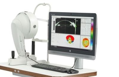
At Omni Eye Centre, our clinic provides advanced diagnostic evaluation and management of keratoconus using modern corneal imaging and specialty contact lenses.
Services offered
- Early keratoconus detection
- Corneal topography interpretation
- Specialty contact lens fitting (including scleral lenses)
- Monitoring before and after corneal cross-linking
- Second opinions for complex cases
Why patients are referred
Many patients are referred when vision cannot be adequately corrected with glasses or standard contact lenses.
2. “Specialty Contact Lens Clinic”
Many eye conditions require advanced contact lenses that go beyond standard soft lenses.
At Omni Eye Centre, we specialise in complex contact lens fittings for patients with irregular corneas and challenging visual needs.
Conditions treated
- keratoconus
- post-surgical corneas
- corneal scarring
- severe dry eye
- contact lens intolerance
3. “Second Opinion Eye Consultation”
If you have ongoing eye problems that have not been resolved, a comprehensive second opinion assessment may help identify the underlying cause.
Patients commonly seek a second opinion for:
- unexplained changes in vision
- persistent contact lens discomfort
- suspected keratoconus
- complications following refractive surgery
Relevant interventions:
- LASIK, Keratoplasty, Hard contact lenses, Scleral Lenses
4. “Eye Rubbing and Corneal Health”
Dr Ared's thesis on eye rubbing is increasingly recognised as a major contributor to corneal changes and as a potential factor in the development of keratoconus.
Our clinic provides specialised assessment for patients with symptoms related to eye rubbing, allergies, and ocular surface disease.
5. “Professional Referrals”
Omni Eye Centre accepts referrals from optometrists, ophthalmologists, and medical practitioners for advanced corneal and contact lens management.
Common referral reasons include:
- keratoconus assessment
- specialty contact lens fitting
- irregular cornea evaluation
- complex dry eye disease
Referring consultants will receive a detailed clinical report following consultation.